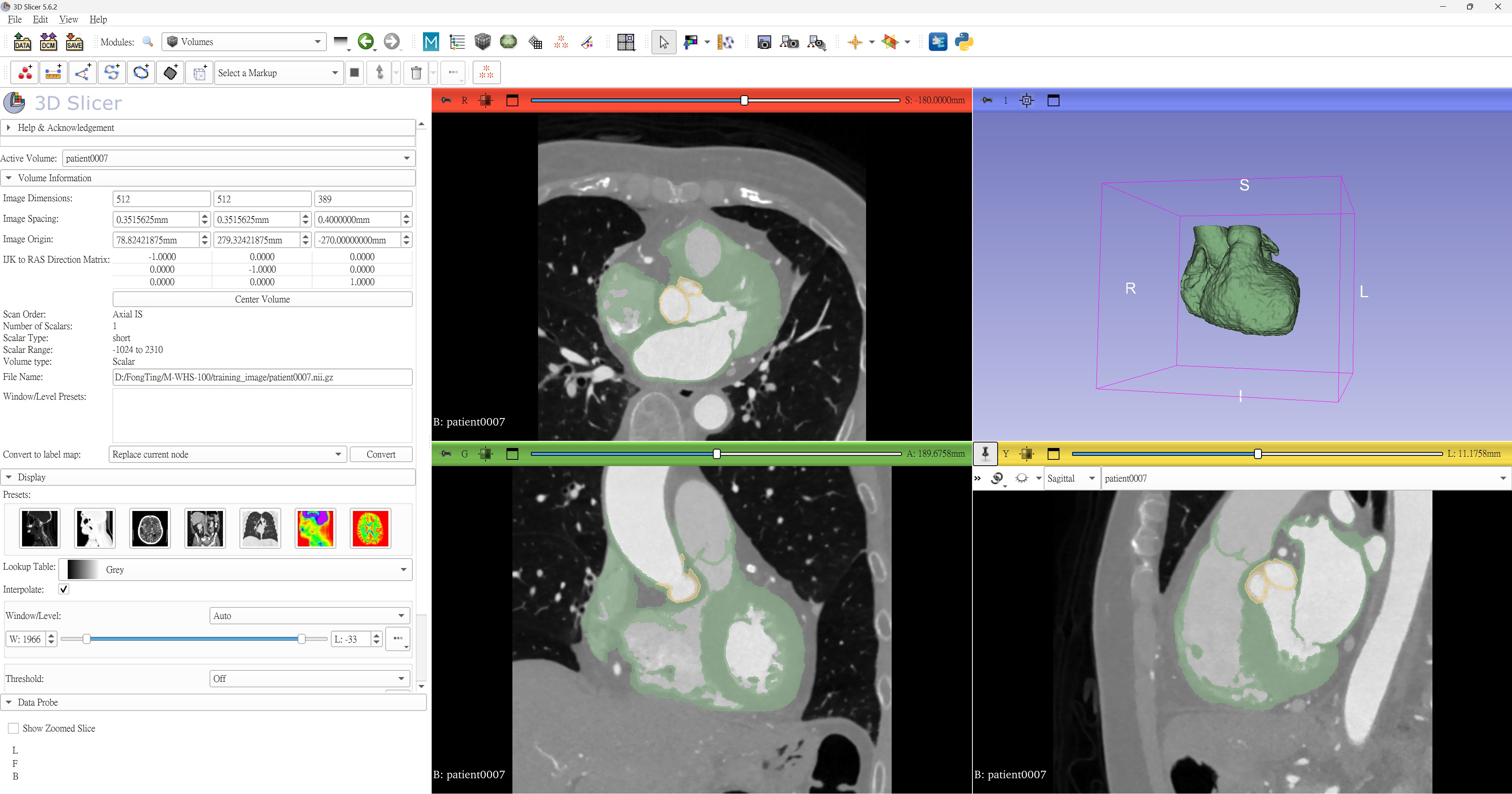
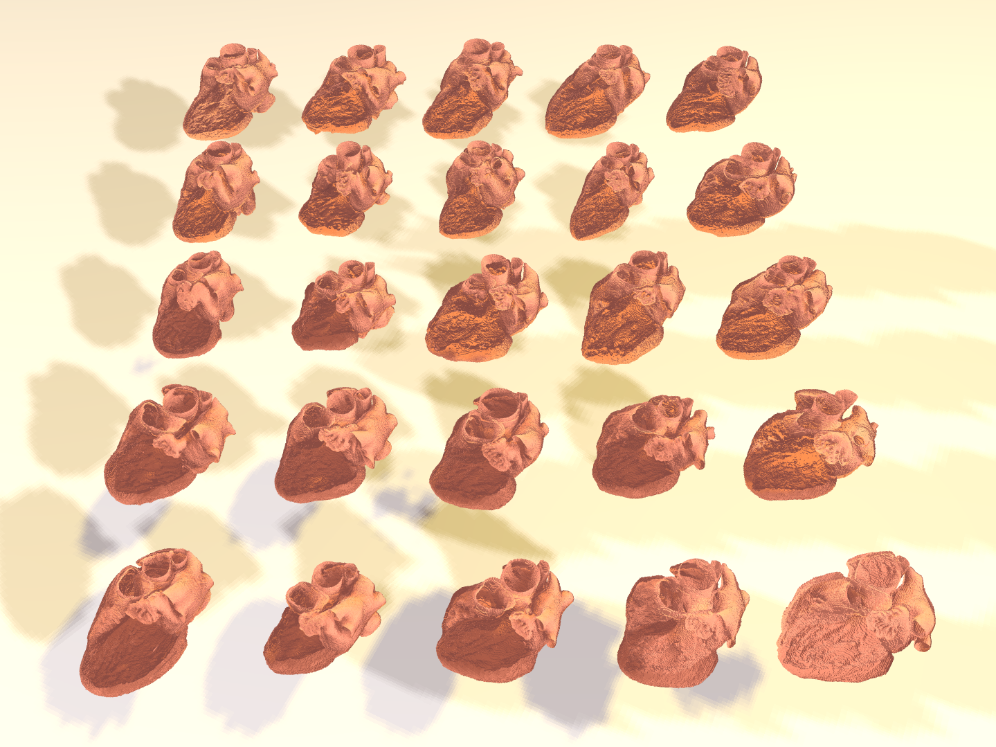
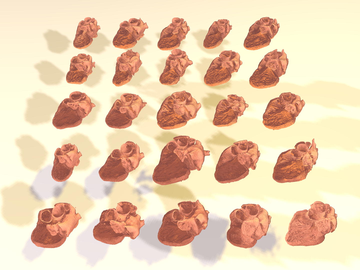
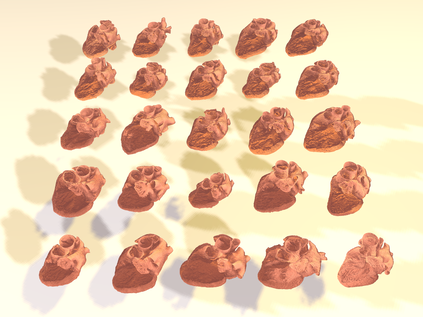
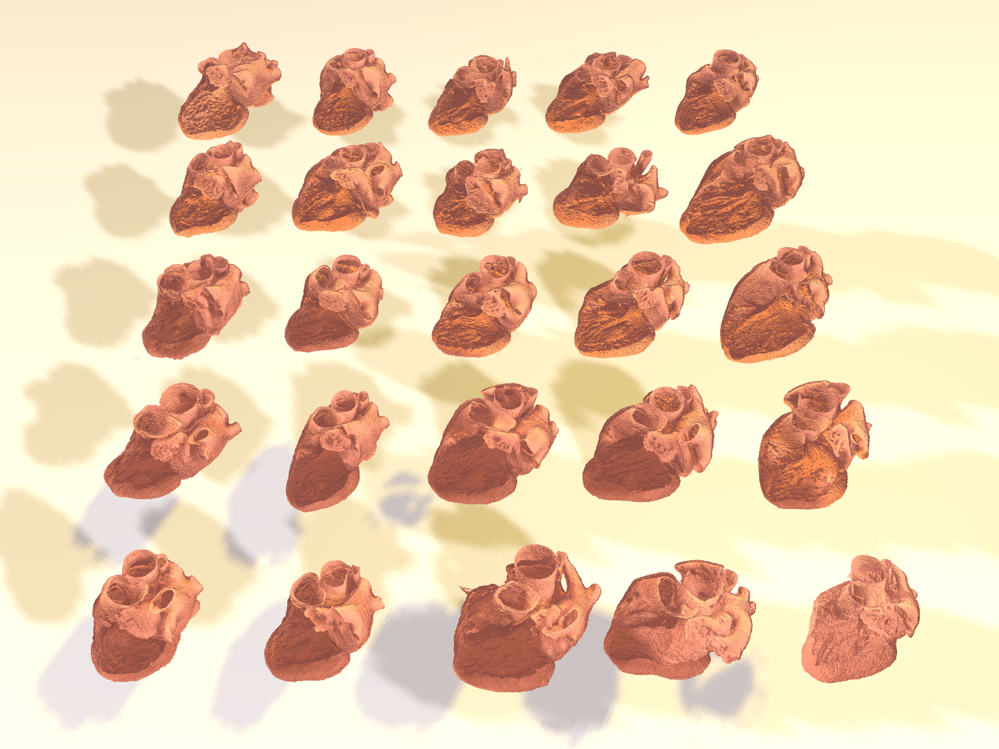
The M-WHS-100 dataset was constructed from clinical CT scans acquired at Cheng Hsin General Hospital. All imaging data were fully anonymized and handled in accordance with the regulations of the hospital's institutional ethics committee. The dataset comprises CT examinations from 100 individual patients and was designed to provide sufficient annotated data for training machine learning-based image segmentation models.
Ground-truth pixel-wise annotations were manually delineated by experts according to a standardized annotation protocol. Segmentation was performed from the basal ring to the sinotubular junction, encompassing the sinus of Valsalva, including the left coronary, right coronary, and noncoronary sinuses, to the maximum extent supported by the available image data. Only the sinus region contiguous with the aortic wall was included, whereas the ascending aorta beyond the sinotubular junction was excluded. Contours were delineated along the luminal boundary of the aortic root while preserving the anatomical boundaries between the left coronary, right coronary, and noncoronary sinuses to ensure accurate separation of the three sinus components.
The resulting ground-truth annotations were encoded as voxel-wise segmentation masks, with label 1 assigned to the myocardium, label 2 to the aortic valve, and label 3 to calcification.